
Challenging Cases
|
|
|
|
|
|
|
|
| What is your diagnosis for these multiple nodules on dorsal tongue? | |
 |
|
| Clinical History | Histologic Examination |
| Please select the most likely answer from the following three choices | |
| (A) Fibroma --- Submit | |
| (B) Amyloidosis --- Submit | |
|
(C) Hemangiolymphangioma ---
Submit
|
|
| Please study carefully the clinical history, panorex findings, and histologic pictures provided below. | |
 |
 |
|
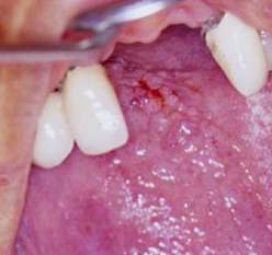
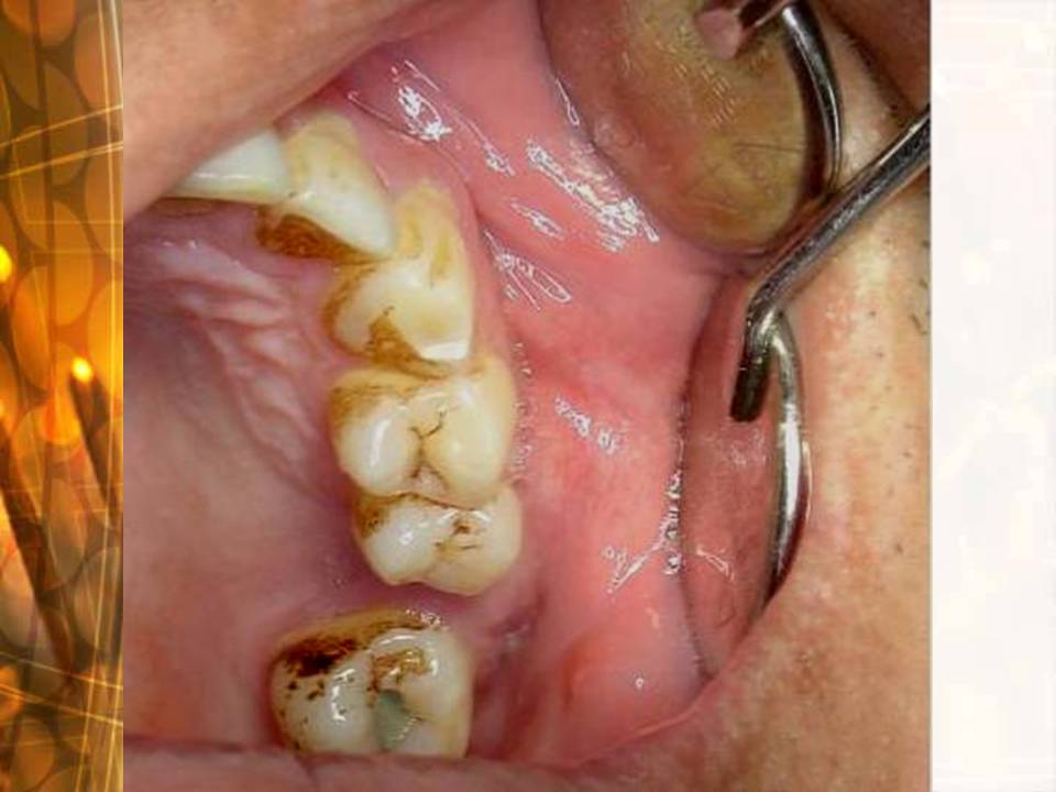
Clinical history (above
left): A 42 y/o female patient complained pain over left mandibular posterior teeth. Oral examination revealed mild buccal & lingual bony expansion over tooth 37 and retromolar area with a normal covering mucosa.
|
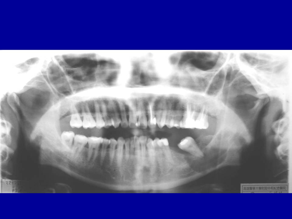
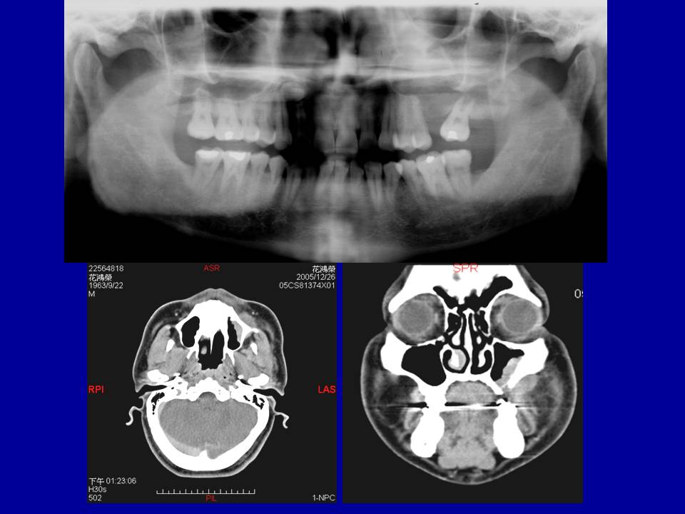
Panorex findings (above
right): There was a well-defined multilocular ovoid-shaped radiolucence with a sclerotic margin around the root of tooth 38 extending from retromolar area down to the inferior border of left mandible with vertical expansion, & from mesial aspect of tooth 38 to left mandibular angle, measuring about 3.0 × 6.0 cm in diameter. |
| Histologic Picture (1) | Histologic Picture (2) |
| Please provide the most likely answers for the following four questions | |
| (1) What is your differential diagnosis for this mandibular lesion? | |
| (2) Does any special stain(s) should be done to confirm your diagnosis? | |
| (3) What is the most likely diagnosis for this mandibular lesion? | |
|
(4)
How to treat this
mandibular lesion?
Answers submitted
|
|
| Please study carefully the clinical history, panorex and CT findings as well as the histologic pictures provided below. | |
 |
 |
|
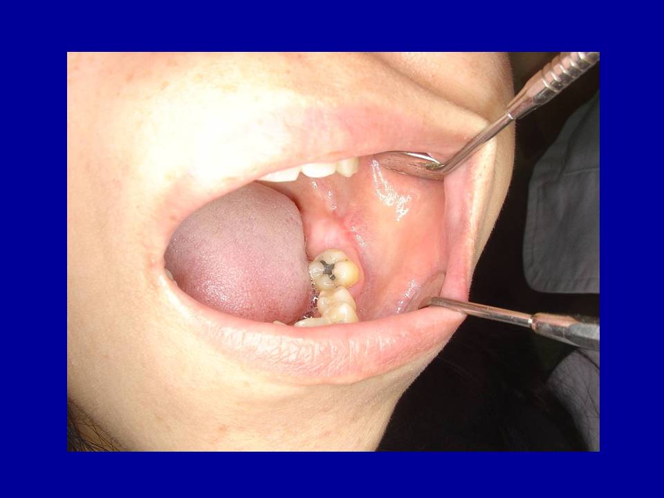
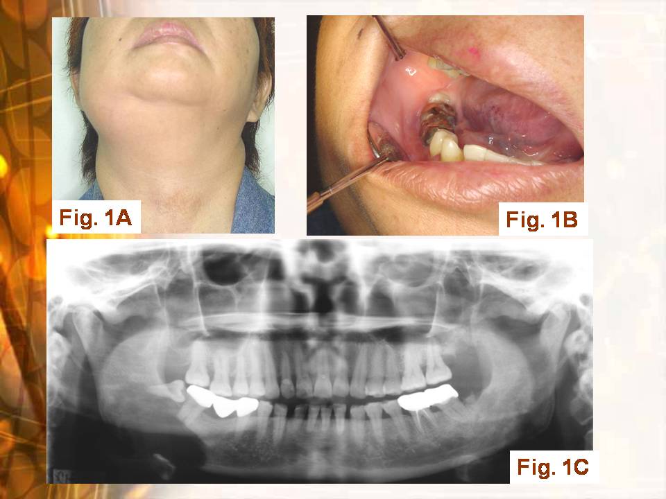
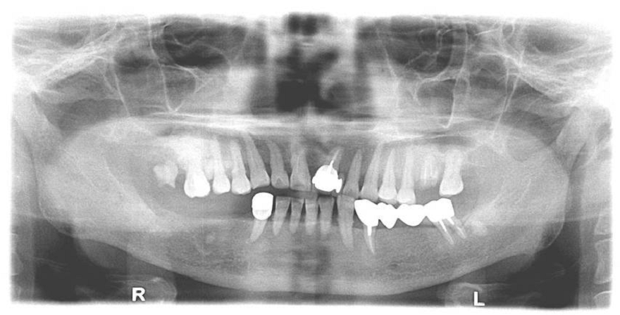
Extraoral findings (Fig.
1A): Swelling over right mandibular body-angle to submandibular area 1. Size: 5×3 cm 2. Consistency: fixed and hard 3. Pain: (+) 4. Paresthesia over right lower lip and gingiva
|
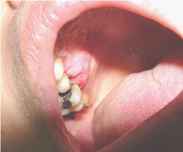
Intraoral findings (Fig. 1B): A swelling mass from teeth 47, 48, to retromolar area with shallow vestibule. 1. Size: 5×2.8 cm 2. Surface: smooth 3. Color: pink 4. Consistency: bony hard 5. Pain: (+) 6. Right lower lip and gingiva paresthesia |
| Histologic Picture (1) | Histologic Picture (2) |
| Please provide the most likely answers for the following four questions | |
| (1) What is your most likely differential diagnosis for this lesion? | |
| (2) Does any special stain(s) should be done to confirm your diagnosis? | |
| (3) How to treat this mandibular lesion? | |
|
(4) What further
examination(s) would you suggest for this patient? Answers submitted |
|
| Please study carefully the clinical history, panorex and CT findings as well as the histologic pictures provided below. | |
 |
 |
| Clinical
history This 43 y/o male suffered from left facial swelling for one month. Intraoral findings
(above left): |
Panorex
finding (above right) An ill-defined radiolucence over left posterior maxilla.
|
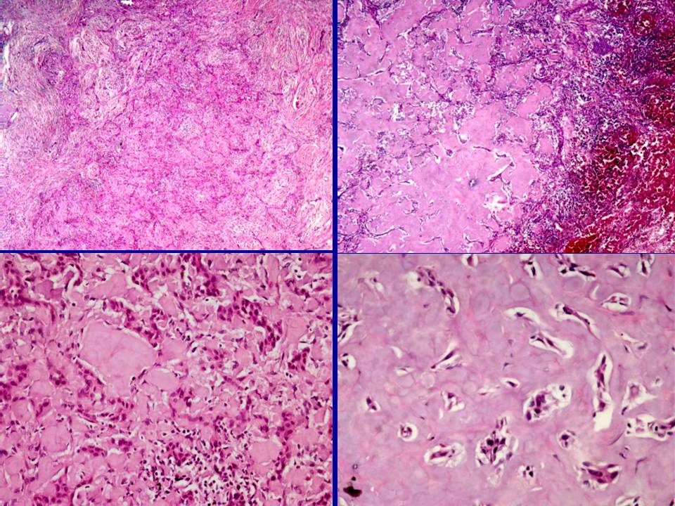
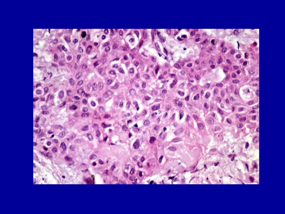
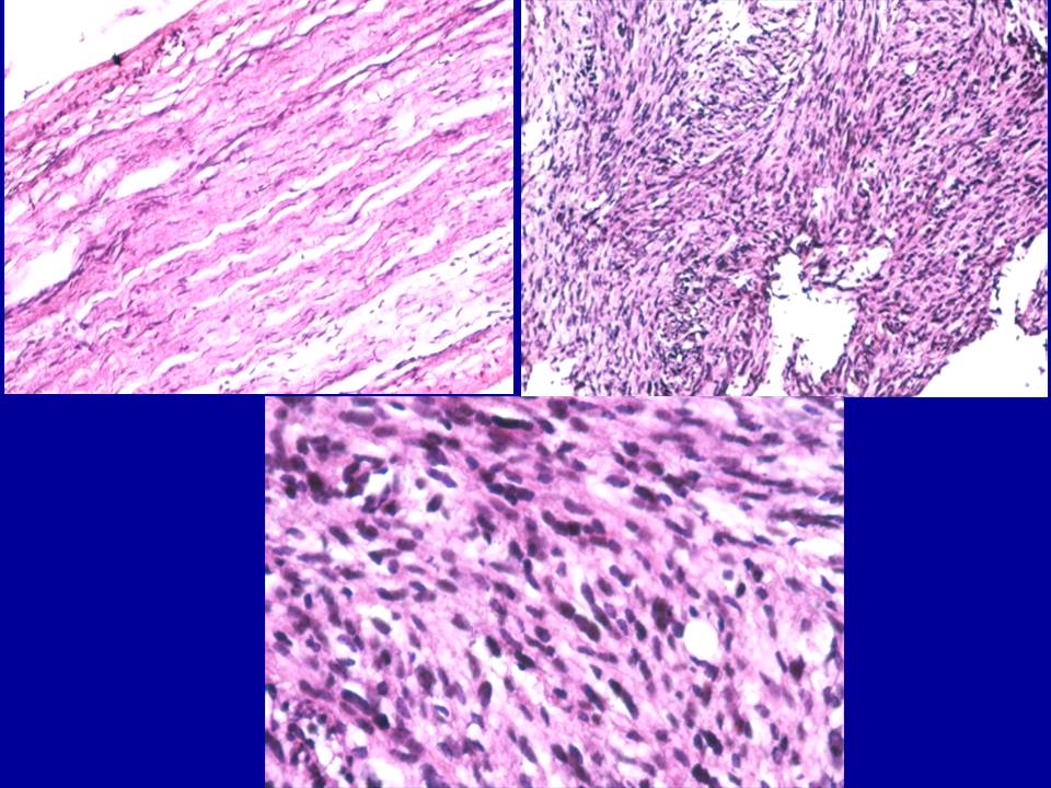
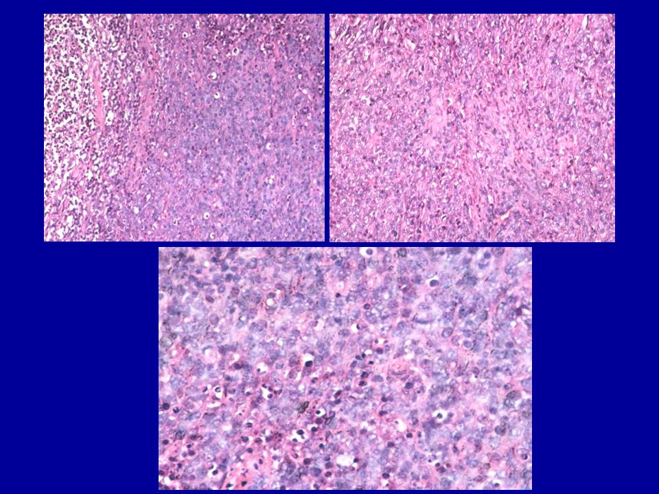
| Histologic Picture | |
| Please provide the most likely answers for the following four questions | |
| (1) What is your differential diagnosis for this lesion? | |
| (2) What is your most likely diagnosis for this lesion? | |
|
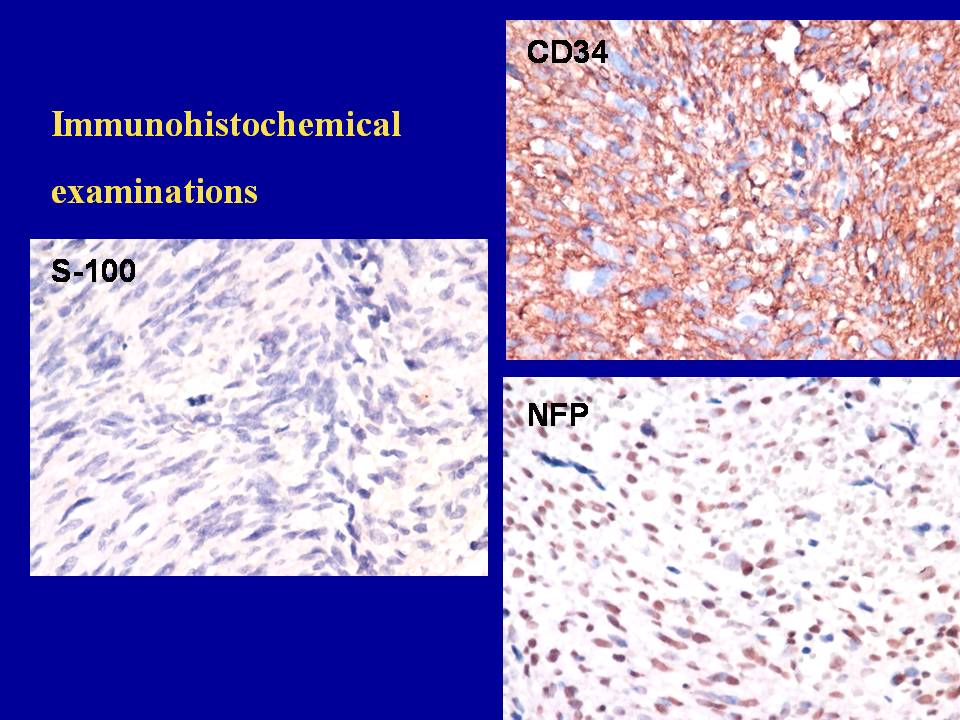
(3) What immunohistochemical
stainings have to be done to confirm the pathologic diagnosis? |
|
|
(4) How to treat this
patient?
Answers submitted
|
|
| Please study carefully the clinical history, panorex and the histologic pictures provided below. | |
 |
 |
|
Clinical
history Intraoral findings
(above left): |
Panorex
finding (above right) A radiopaque (apical 14-16) mixed with radioluce (17-tuberosity) lesion extended from tooth 14 apical to tuberosity from alveolar crest to sinus floor, measured about 4.5×2.0 cm in diameter. |
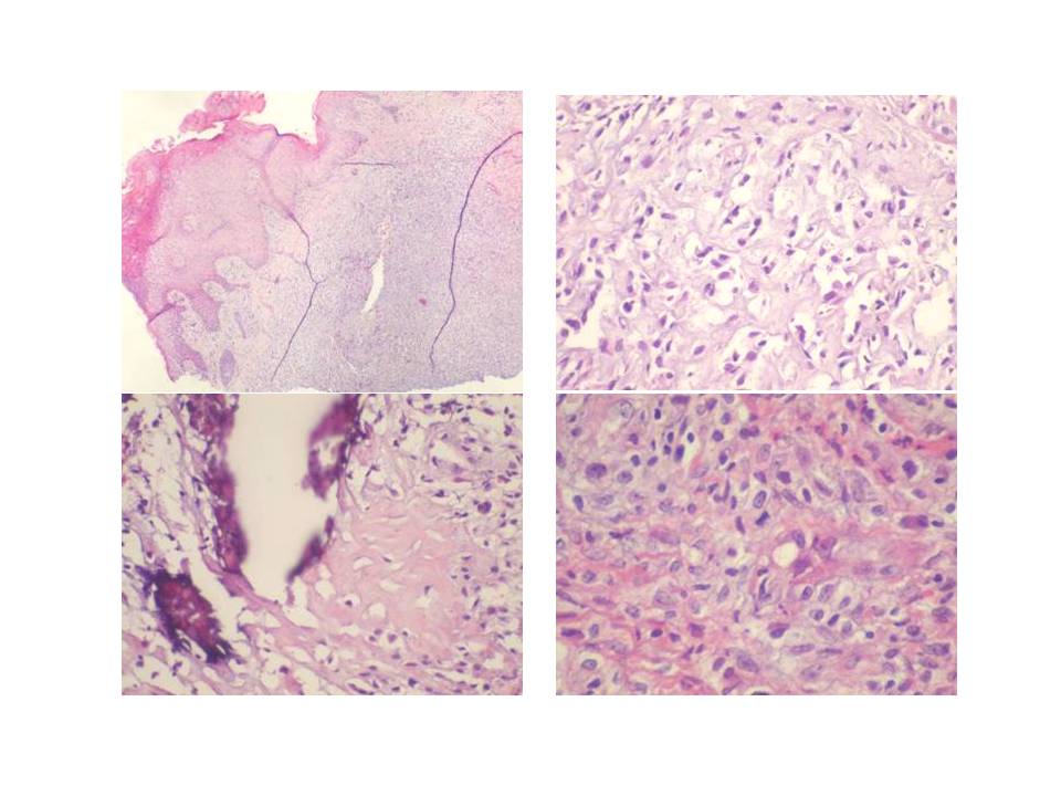
| Histologic Pictures | |
| Please provide the most likely answers for the following four questions | |
| (1) What is your differential diagnosis for this lesion? | |
| (2) What is your most likely diagnosis for this lesion? | |
|
(3) What immunohistochemical
stainings have to be done to confirm the pathologic diagnosis? |
|
|
(4) How to treat this
patient?
Answers submitted
|
|
![]()